Research Facilities
Siemens 3T Vida scanner with a 64-channel head coil
(Neuroimaging research is migrating to this new facility at the MSU Health Care at McLaren Greater Lansing Outpatient Imaging Center)
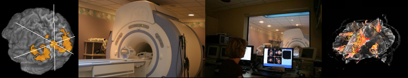
GE 3T Signa® HDx MR scanner (retired)
(Room E120, Radiology Building)
This system has an actively shielded short-bore (2.06 m width x 1.72 m length) magnet, a fast gradient system that provides high-speed brain imaging for fMRI as well as regular body imaging, a data acquisition system supporting up to 32 channels, an 8-channel head coil and other coils for this platform, a powerful volume reconstruction engine that enables virtually real-time image generation even when massive parallel imaging datasets are involved, as well as multinuclear spectroscopy hardware. The short-bore magnet provides a low incidence of claustrophobia. The gradient rise time (150 T/m/s) and peak gradient strength (50 mT/m per axis) are among the best in the industry for whole-body systems. Parallel imaging technology allows EPI images to be acquired with higher temporal resolution and with less distortion for fMRI studies and other EPI-based image acquisitions. This MRI system will also be capable of running fMRI studies in a "real-time" mode, in which an investigator can assess the data quality during scanning, and see the activation maps after each functional run. MRI research at MSU also benefits from a close collaborative relationship and formal research agreement with GE. All technical aspects of the scanner are accessible to MRI physicists, including the pulse programming module (EPIC).
MR-compatible eye tracker
The CIRC has an MR-compatible eye tracker, the Eyelink 1000 Plus from SR research (Ottawa, Canada). This system has high spatial and temporal resolution and can be used for accurate tracking of subjects' eye position during the imaging session. More technical specifications of the system can be found on the manufacturer's websit. Interested users should contact Dr. Jan Brascamp for more usage-related information.Other equipment
The CIRC has a high-performance PC equipped with E-Prime (Psychology Software Tools, Inc., Pittsburgh, PA) and Matlab to control visual and auditory stimulus presentation and to record participant responses. The computers are used to control audio and or video playback in synchronization with MRI scan timing. A 1024 x 768 32-inch LCD monitor with a 10 x 13 degree of visual angle (Salvagione Design, Sausalito, CA) and the MR-compatible Hyperion digital projection system (resolutions: default at 1024 x 768 and native at 1920 x 1080) (Psychology Software Tools, Inc., Pittsburgh, PA) with a 23 x 30 degree of visual angle placed at the back of the magnet room are available for presenting visual stimulus in fMRI studies. The Serene Sound Audio System (Resonance Technology Inc, Northridge, CA) is available for studies requiring auditory stimuli. A BrainLogics Fiber Optic Button Response System with a pair of 5-button MR-compatible keypads (Psychology Software Tools, Inc., Pittsburgh, PA) and an MR-compatible mouse (Mag Design and Engineering, Redwood City, CA) are used to record participant responses. An MR-compatible small-size “12M-i” camera with integrated LED infrared light (MRC Systems GmbH, Germany) is available to monitor face and eye movement. A Persaio MRI Noise Cancellation System (Psychology Software Tools, Inc., Pittsburgh, PA) is available to hear and record clear subject verbal response. In addition, a mock scanner is available. This device presents the look, feel and sound of our MRI scanner. Exposure to the mock scanner prior to actual scanning helps subjects habituate to the scanning environment.
Data and Image processing
The CIRC has two dedicated Linux servers for the storage of image data, where investigators can retrieve their data. The data is also routinely backed up to a tape drive. CIRC also has a few Linux workstations and PCs for investigators and students to process data and prepare fMRI stimulus paradigms.